 Fundus fluorescein angiography is a medical procedure in which a fluorescent dye is injected into the bloodstream. The dye highlights the blood vessels in the back of the eye so they can be photographed.
This test is often used to manage eye disorders. To confirm a diagnosis, determine an appropriate treatment, or monitor the condition of the vessels in the back of your eye.
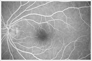
In FFA, the chemical dye is injected in the vein of the hand and this dye circulates all over the body and in every blood vessel. Since retina is the only place in the body where blood vessels can be seen, it is used to visualize the retinal vasculature that is blood vessels of the retina. A special machine called the fundus camera, which has got special lens filters, is used to take photographs of the retina. We can also attach digital camera system or a printer. When blue light falls on retina which has Fluorescein dye the reflected light is of green colour & not blue. This property of dye is used to study the normal or abnormal anatomy of retina. This dye and the reflected light are greenish in color. The fundus camera records this. This dye would go wherever the blood vessels are present in retina and we would get the picture of that.
If blood vasculature of the retina is normal, then we would get the normal pattern of the retina. If there were blockage then the dye would not go beyond it and would stop at that particular point. If there is leakage, the dye would also leak out along with the blood in the retina and we would be able to pick up the leakage. If there are certain other defects in the retina then depending on the various features seen, whether we get leakages or staining or blocked fluorescence or window defects whether the dye seen in early phase, late phase or in very late phase or delayed phase and is initially less and increasing etc, we can get very good idea of patients disease.